
NOAA Teacher at Sea
Dieuwertje “DJ” Kast
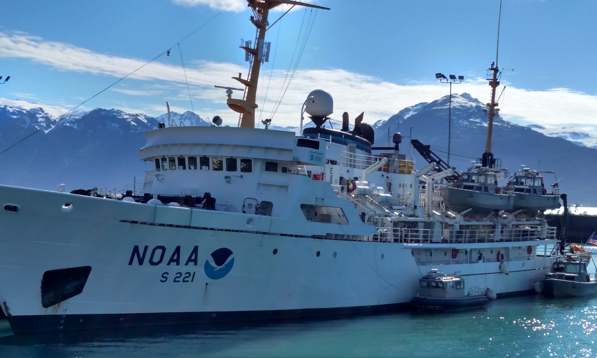
Aboard NOAA Ship Henry B. Bigelow
May 19 – June 3, 2015
Mission: Ecosystem Monitoring Survey
Geographical area of cruise: Gulf of Maine
Date: May 28, 2015, Day 11 of Voyage
Interview with Student Megan Switzer

Megan Switzer is a Masters student at the University of Maine in Orono. She works in Dave Townsend’s lab in the oceanography department. Her research focuses on interannual nutrient dynamics in the Gulf of Maine. On this research cruise, she is collecting water samples from Gulf of Maine, as well as from Georges Bank, Southern New England (SNE), and the Mid Atlantic Bight (MAB). She is examining the relationship between dissolved nutrients (like nitrate and silicate) and phytoplankton blooms. This is Megan’s first research cruise!
In the generic ocean food chain, phytoplankton are the primary producers because they photosynthesize. They equate to plants on land. Zooplankton are the primary consumers because they eat the phytoplankton. There are so many of both kinds in the ocean. Megan is focusing on a particular phytoplankton called a diatom; it is the most common type of phytoplankton found in our oceans and is estimated to contribute up to 45% of the total oceanic primary production (Yool & Tyrrel 2003). Diatoms are unicellular for the most part, and a unique feature of diatom cells is that they are enclosed within a cell wall made of silica called a frustule.


The frustules are almost bilaterally symmetrical which is why they are called di (2)-atoms. Diatoms are microscopic and they are approximately 2 microns to about 500 microns (0.5 mm) in length, or about the width of a human hair. The most common species of diatoms are: Pseudonitzchia, Chaetocerous, Rhizosolenia, Thalassiosira, Coschinodiscus and Navicula.


Diatoms also have ranges and tolerances for environmental variables, including nutrient concentration, suspended sediment, and flow regime. As a result, diatoms are used extensively in environmental assessment and monitoring. Furthermore, because the silica cell walls are inorganic substances that take a long time to dissolve, diatoms in marine and lake sediments can be used to interpret conditions in the past.
In the Gulf of Maine, the seafloor sediment is constantly being re-suspended by tidal currents, bottom trawling, and storm events, and throughout most of the region there is a layer of re-suspended sediment at the bottom called the Bottom Nepheloid Layer. This layer is approximately 5-30 meters thick, and this can be identified with light attenuation and turbidity data. Megan uses a transmissometer, which is an instrument that tells her how clear the water is by measuring how much light can pass through it. Light attenuation, or the degree to which a beam of light is absorbed by stuff in the water, sharply increases within the bottom nepheloid layer since there are a lot more particles there to block the path of the light. She also takes a water sample from the Benthic Nepheloid Layer to take back to the lab.

Megan also uses a fluorometer to measure the turbidity at various depths. Turbidity is a measure of how cloudy the water is. The water gets cloudy when sediment gets stirred up into it. A fluorometer measures the degree to which light is reflected and scattered by suspended particles in the water. Taken together, the data from the fluorometer and the transmissometer will help Megan determine the amount of suspended particulate material at each station. She also takes a water sample from the Benthic Nepheloid layer to take back to the lab. There, she can analyze the suspended particles and determine how many of them are made out of the silica based frustules of sinking diatoms.

She collects water at depth on each of the CTD/ Rosette casts.


CTD, Rosette, and Niskin Bottle basics.
The CTD or (conductivity, temperature, and depth) is an instrument that contains a cluster of sensors, which measure conductivity, temperature, and pressure/ depth.
Here is a video of a CTD being retrieved.
Depth measurements are derived from measurement of hydrostatic pressure, and salinity is measured from electrical conductivity. Sensors are arranged inside a metal housing, the metal used for the housing determining the depth to which the CTD can be lowered. Other sensors may be added to the cluster, including some that measure chemical or biological parameters, such as dissolved oxygen and chlorophyll fluorescence. Chlorophyll fluorescence measures how many microscopic photosynthetic organisms (phytoplankton) are in the water. The most commonly used water sampler is known as a rosette. It is a framework with 12 to 36 sampling Niskin bottles (typically ranging from 1.7- to 30-liter capacity) clustered around a central cylinder, where a CTD or other sensor package can be attached. The Niskin bottle is actually a tube, which is usually plastic to minimize contamination of the sample, and open to the water at both ends. It has a vent knob that can be opened to drain the water sample from a spigot on the bottom of the tube to remove the water sample. The scientists all rinse their bottles three times and wear nitrile or nitrogen free gloves to prevent contamination to the samples.
On NOAA ship Henry B. Bigelow the rosette is deployed from the starboard deck, from a section called the side sampling station of this research vessel.
The instrument is lowered into the water with a winch operated by either Adrian (Chief Boatswain- in charge of deck department) or John (Lead Fishermen- second in command of deck department). When the CTD/Rosette is lowered into the water it is called the downcast and it will travel to a determined depth or to a few meters above the ocean floor. There is a conducting wire cable is attached to the CTD frame connecting the CTD to an on board computer in the dry lab, and it allows instantaneous uploading and real time visualization of the collected data on the computer screen.

The water column profile of the downcast is used to determine the depths at which the rosette will be stopped on its way back to the surface (the upcast) to collect the water samples using the attached bottles.
Niskin Bottles:
Messenger- The manual way to trigger the bottle is with a weight called a messenger. This is sent down a wire to a bottle at depth and hits a trigger button. The trigger is connected to two lanyards attached to caps on both ends of the bottle. When the messenger hits the trigger, elastic tubing inside the bottle closes it at the specified depth.


Here is a video of how the manual niskin bottle closes: https://www.youtube.com/watch?v=qrqXWtbUc74
The other way to trigger Niskin bottles is electronically. The same mechanism is in place but an electronic signal is sent down the wire through insulated and conductive sea cables (to prevent salt from interfering with conductivity) to trigger the mechanism.
Here is a video of how it closes electronically: https://www.youtube.com/watch?v=YJF1QVe6AK8


Using the Niskin bottles, Megan collects water samples at various depths. She then filters water samples for her small bottles with a syringe and a filter and the filter takes out the phytoplankton, zooplankton and any particulate matter. She does this so that there is nothing living in the water sample, because if there is there will be respiration and it will change the nutrient content of the water sample.


This is part of the reason why we freeze the sample in the -80 C fridge right after they have been processed so that bacteria decomposing can’t change the nutrient content either.
Diatoms dominate the spring phytoplankton bloom in the Gulf of Maine. They take up nitrate and silicate in roughly equal proportions, but both nutrients vary in concentrations from year to year. Silicate is almost always the limiting nutrient for diatom production in this region (Townsend et. al., 2010). Diatoms cannot grow without silicate, so when this nutrient is used up, diatom production comes to a halt. The deep offshore waters that supply the greatest source of dissolved nutrients to the Gulf of Maine are richer in nitrate than silicate, which means that silicate will be used up first by the diatoms with some nitrate left over. The amount of nitrate left over each year will affect the species composition of the other kinds of phytoplankton in the area (Townsend et. al., 2010).
The silica in the frustules of the diatom are hard to breakdown and consequently these structures are likely to sink out of the euphotic zone and down to the seafloor before dissolving. If they get buried on the seafloor, then the silicate is taken out of the system. If they dissolve, then the dissolved silicate here might be a source of silicate to new production if it fluxes back to the top of the water column where the phytoplankton grow.
Below are five images called depth slices. These indicate the silicate concentration (rainbow gradient) over a geographical area (Gulf of Maine) with depth (in meters) latitude and longitude on the x and y axis.

This is the type of data Megan is hoping to process from this cruise.
Very Informative and definitely a good read.